- Diagnositc Kits
- MAR Diagnostic Kit for Anti-Sperm Antibody
- Semen Leukocyte Peroxidase Staining Kit
- One-time Semen processing Kit
- Sperm Hypo-Osmotic Staining Kit
- Sperm Life Detection Kit (Eosin Staining Method)
- Sperm Staining Detection Kit (Rapid Staining Metho
- Sperm Acrosin Detection Kit (PSA-FITC Staining Met
- Sperm DNA Fragmentation Staining Kit (Wright-Giems
- Diagnostic Kit for Leptin (ELISA)
- Immunohistochemical Detection Kit for P185erbB-2/


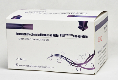
Immunohistochemical Detection Kit for P185erbB-2/HER-2 Oncoprotein
SPECIFICATION AND INSTRUCTION
[Product Name]
Immunohistochemical Detection Kit for P185erbB-2/HER-2 Oncoprotein
[Package]
20 samples per kit
[Intended Use]
For detecting tumor protein p185erbB-2/HER-2 provide the basis of diagnosis in Breast cancer and ovarian cancer .
[Test Principle]
erbB-2/HER-2 is a oncogene, the protein of 185kD(p185),the positive expression of p185 becomed a significant tumor marker of international generally accepted. This kit uses self-developed Anti-p185erbB-2/HER-2 Monoclonal antibody, and perform Biotin-Avidin IHC , cancer membrane Stained yellow means positive .
[Units of Kit]
(1) Solution A 5.0ml 3% H2O2
(2) Solution B 1.2ml Blocking Solution
(3) Solution C 1.2ml Anti-p185erbB-2/HER-2Monoclonal antibody
(4) Solution D 1.2ml Biotinylated secondary antibody
(5) Solution E 1.2ml Avidin-labeled HRP
(6) DAB prepared before use
(7) positive tissue slice (Purchased youself) : tissue slice of p185erbB-2/HER-2 positive
(8) Instruction.
[Storage]
Stored at 2 ~ 8℃ for 10 months
[Equipment]
Common light microscope
[Sample]
Tissue slice
[Test method]
(1) Paraffin Sections: Trim paraffin blocks as necessary and cut at 4 um (5 um is commonly used).And the frozen sections,should have been used fixation for frozen tissue sections is to immerse the slides in pre-cooled acetone 4℃ for 10 min;
(2) Dry sections, melting Paraffin:Dry sections in incubator for at least 15 min. at -90℃;
(3)Deparaffinization/Rehydration:Hydrate with Xylene thanol and rinse in distilled water,then can perform Immunohistochemistry;
(4) Rinse in PBSH7.4,0.01M Phosphate buffer or 3x5min;
(5) Allow the slide to drain, Shake off excess Liquid with a brisk motion and carefully wipe each slide around the Sections, Dropping A solution cover the tissue Sections ,incubate
10 min at Room temperature. Shake off excess Liquid, Rinse in PBS for 3x5min;
(6) Allow the slide to drain, Shake off excess Liquid with a brisk motion and carefully wipe each slide as before ,apply B solution cover the tissue Sections ,incubate 10 min at Room temperature;
(7) Allow the slide to drain, Shake off excess Liquid with a brisk motion and carefully wipe each slide as before, apply C solution cover the tissue Sections, apply B solution cover the tissue Section. ,incubate for 1 hour at 37℃ or overnight at 4℃chamber. Rinse in PBS for 3x5min; Allow the slide to drain, Shake off excess Liquid with a brisk motion and carefully wipe each slide as before, apply B solution cover the tissue Section, incubate 10 min at Room temperature, Rinse in PBS for 3x5min;
(8) Allow the slide to drain, Shake off excess Liquid with a brisk motion and carefully wipe each slide as before ,apply E solution cover the tissue Sections ,incubate 10 min at Room temperature, Rinse in PBS for 3x5min;.
(9) Prepared DAB solution when it been used, dissolve 5mgDAB in 10ml of PBS,and apply A solution 100µl, (uint desired color reaction is observed when monitored with the Microscope);
(10) Rinse in distilled water ,or can do karyotin again when necessary; Dehydrate through Gradient ethanol, Clear in xylene, Coverslip with Gum.
[Reference Result]
Reference value: compared to negative tissue slice ,the positive tissue slice should have 5% cancer membrane Stained yellow, immune reactions were continuous and linear around cell.
[Interpretation of the Results Identified]
Negative: the tissue Sections without stained, <5% cancer membrane Stained yellow, Staining intensity is Low.
Low positive (±-+) :> 5% cancer membrane Stained yellow, immune reactions were not continuous and linear around cell . Staining intensity is Low,part of cell Stained.
Moderate positive(++): > 5% cancer membrane Stained yellow, immune reactions were continuous and linear around cell
High positive(+++): > 5% cancer membrane Stained dense yellow, immune reactions were continuous and linear around cell.++-+++ means over-expression
Precaution: The stained Cytoplasm can not be judged for Specific staining .
[Limitations of Test Methods]
Only for tissue slice
[Product Performance]
Comparison with similar products, Correlation coefficient0.654<0.0001
[Precaution]
(1) By use this kit,the Sections without prior enzyme digestion or antigen rerival.
(2) All solutions applied should cover the tissue Sections
(3) Room temperature mean 20 �, Bath at 37 � better
(4) pH7.4, 0.01MPBS and DAB need Prepared before use by youself
[Manufactured by]
Anhui Anke Biotechnology (Group) Co., Ltd.
[Address]
Ankebio Buildings, 669, Changjiang Road, West ,Hefei, 230088, P.R. China.
Tel: +86-551-65319890, 65318811
Fax:+86-551-65319895
Website: www.ankebio.com
E-mail: info@ankebio.com
[Medical Device Manufacturing Enterprise License Number] WSYJX 20070120
[The registration Number of Medical Devices] GSYJX(Z)Z2009N3400543
[Standard Number] YBS00392004
[Instruction Approval and Modified day] July 21st ,2009

